
AHI PedCAT CASE STUDIES
CASE 1 -- TARSAL COALITION
Clinical History
25-year-old female who sustained an MVA 14 months ago injuring the right ankle, status post ORIF for bimalleolar fractures, complaining of persistent right ankle pain and reduced range of motion.
Plain Films
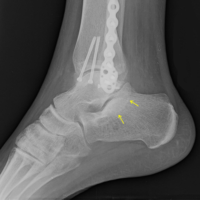
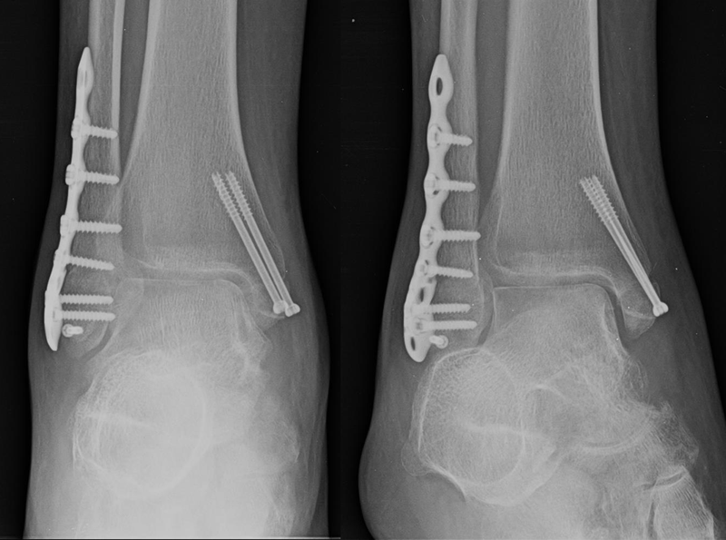
Lateral, AP and Oblique views of the right ankle show orthopedic hardware and healed bimalleolar fractures. No tarsal coalition is demonstrated. The posteroinferior aspect of the sustentaculum tali appears somewhat prominent (arrows).
PedCAT Scan
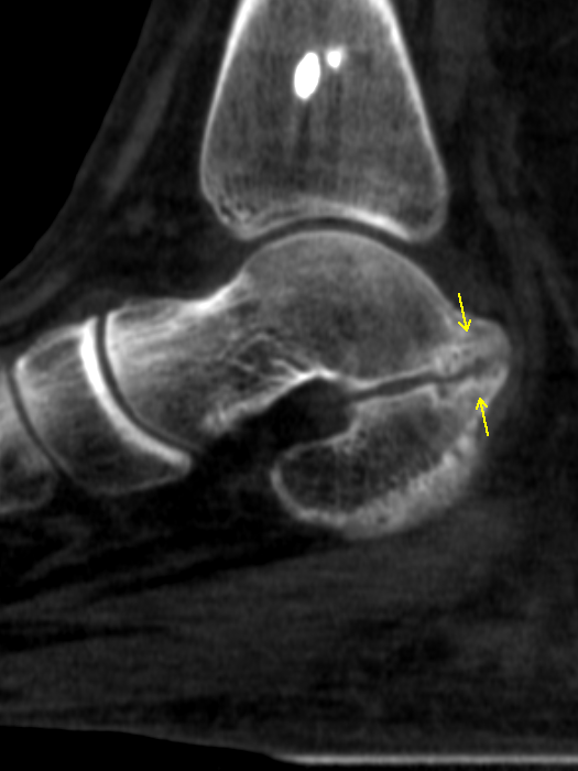
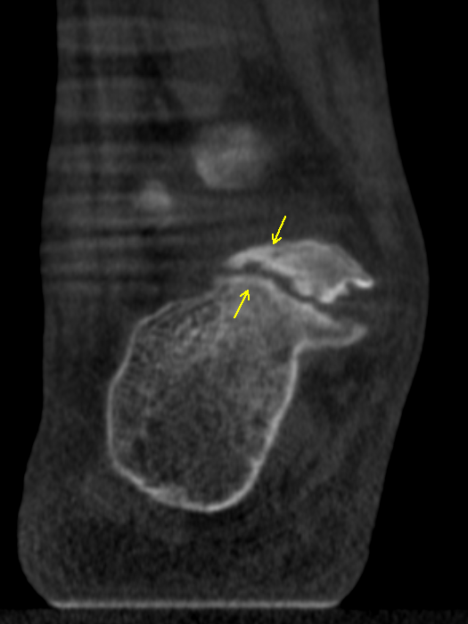
Sagittal and coronal sections from the PedCAT scan clearly demonstrate non-osseous talocalcaneal coalition involving the posterior subtalar joint, with irregularity of the articular surfaces, narrowing of the joint space, and subchondral sclerosis (arrows).
PedCAT Impact
The PedCAT scan quickly and easily estblished the diagnosis of talocalcaneal coalition, and outlined the type, size, location and extent of the coalition.
CASE 2 -- SUBTALAR ARTHRODESIS
Clinical History
65-year-old male status post subtalar arthrodesis one year ago, complaining of persistent right ankle pain.
Plain Films
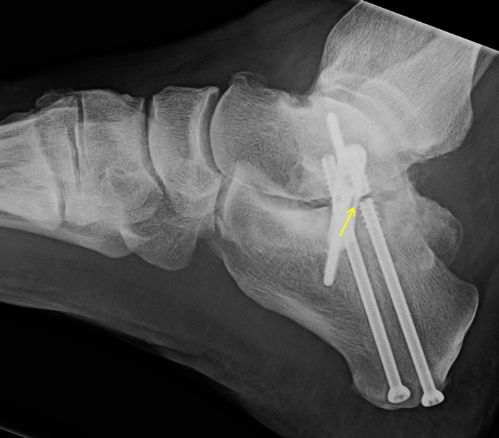
Lateral, Oblique and DP views of the right foot show a fracture in the more posterior screw through the posterior subtalar joint (arrow). The other screws appear intact. No bony fusion can be seen.
PedCAT Scan
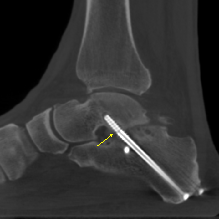
Sagittal and coronal sections from the PedCAT scan demonstrate additional fractures in the more anterior screw through the posterior subtalar joint (arrow), and in a calcaneal screw connected to a small lateral plate (double arrow). Bony fusion is also demonstrated in the lateral aspect of the subtalar arthrodesis (open arrow).
PedCAT Impact
The PedCAT scan quickly and easily estblished the lack of integrity of several harware components, and the presence and extent of bony fusion in the arthrodesis. The PedCAT scan can also assess for loosenin of the hardware.
CASE 3 -- MIDFOOT FRACTURES
Clinical History
Lorem ipsum dolor sit amet, consectetur adipiscing elit, sed do eiusmod tempor incididunt ut labore et dolore magna aliqua. Ut enim ad minim veniam, quis nostrud exercitation ullamco laboris nisi ut aliquip ex ea commodo consequat.
Plain Films

Sed ut perspiciatis unde omnis iste natus error sit voluptatem accusantium doloremque laudantium, totam rem aperiam, eaque ipsa quae ab illo inventore veritatis et quasi architecto beatae vitae dicta sunt explicabo.
PedCAT Scan

Nemo enim ipsam voluptatem quia voluptas sit aspernatur aut odit aut fugit, sed quia consequuntur magni dolores eos qui ratione voluptatem sequi nesciunt.
PedCAT Impact
Neque porro quisquam est, qui dolorem ipsum quia dolor sit amet, consectetur, adipisci velit, sed quia non numquam eius modi tempora incidunt ut labore et dolore magnam aliquam quaerat voluptatem.
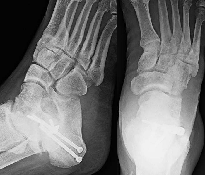
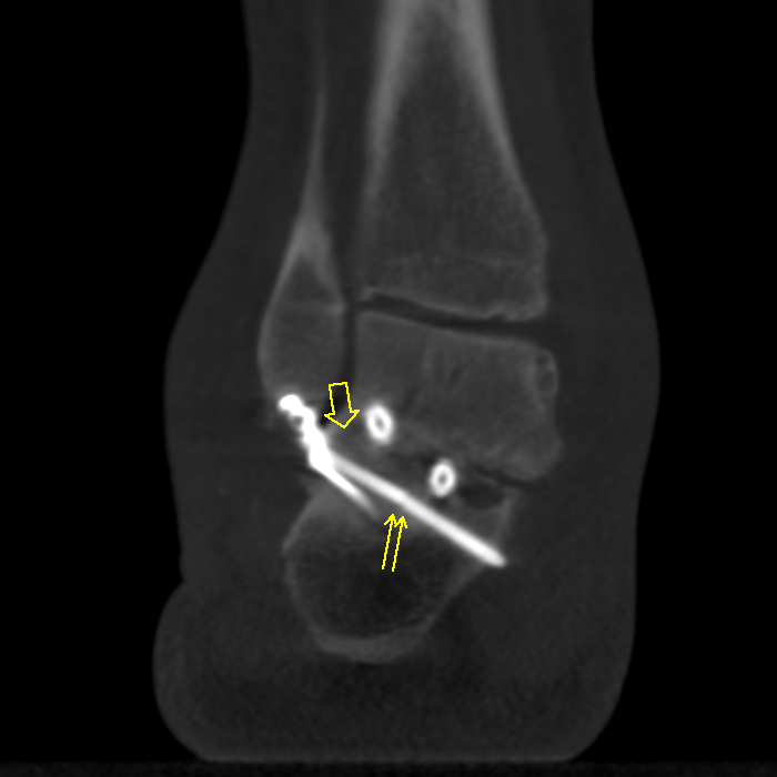
CASE 4 -- SUBTALAR ARTHRODESIS
Clinical History
70-year-old male status post subtalar arthrodesis in the right foot one year ago, complaining of ongoing right foot pain.
Plain Films

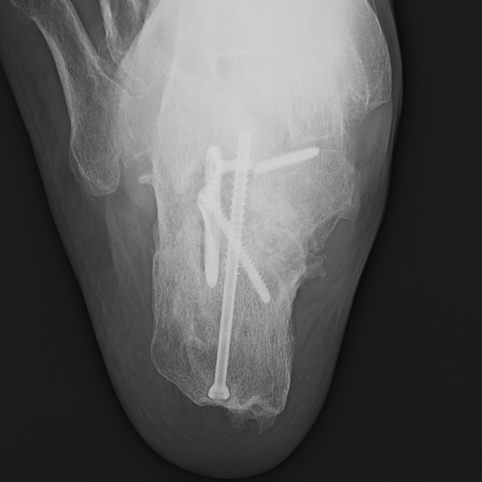
Lateral and Axial views of the right hindfoot show a fracture of one orthopedic screws. The plain films are indeterminate for bony fusion.
PedCAT Scan
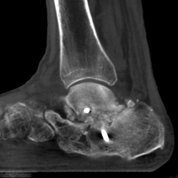
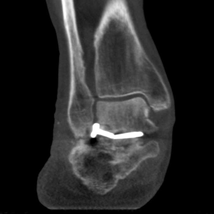
Sagittal and coronal sections from the PedCAT scan again show the fractured screw. The PedCAT scan demonstrates that there is no bony fusion of the arthrodesis.
PedCAT Impact
The PedCAT scan quickly and easily estblished the absence of bony fusion across the arthrodesis. The scan also accurately assessed the integrity of the hardware components.