
AHI MSK CT Protocols
Shoulder CT Protocol
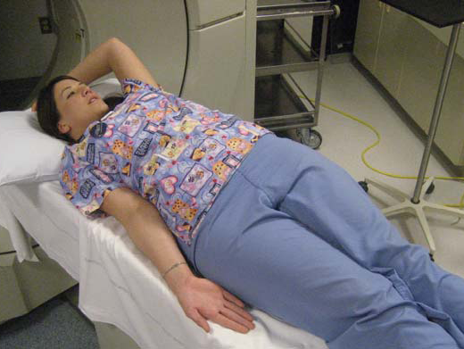
Patient Position
- Supine to mild RPO (keep arm/humerus at approx. mid-coronal plane of body)
- Affected arm by side of body with palm of hand facing up (shoulder externally rotated)
- Contralateral arm raised above head
Scan Parameters
- SFOV: Large
- kV: 140
- mAs: 200
Reconstruct
- 1.25/0.62 mm Bone
- 2/2 mm Soft Tissue
Coverage
- From above AC joint to the bottom of the scapula. If there is a shoulder prosthesis, scan to include the distal end of the humeral component.
DFOV
- Just wide enough to include entire scapula and proximal humerus.
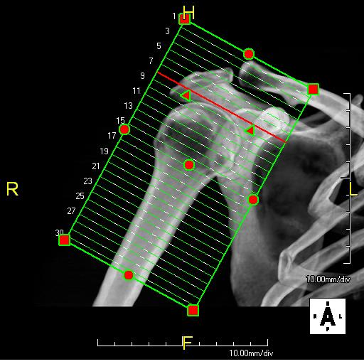
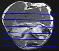
Axial Reformats
- Perpendicular to humeral diaphysis
- 0.8/1.5 mm Bone
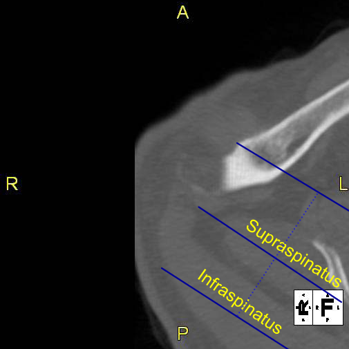
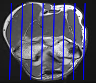
Coronal Reformats
- Prescribe coronal plane off of axial image parallel to supraspinatus muscle.
- 0.8/1.5 mm Bone
Sagittal Reformats
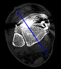
- Prescribe sagittal plane off of axial image perpendicular to mid-glenoid.
- 0.8/1.5 mm Bone
Send Only These...
- Scouts
- Source Bone Reconstructions
- Source Soft Tissue Reconstructions
- Axial Reformats
- Coronal Reformats
- Sagittal Reformats
Elbow CT Protocol
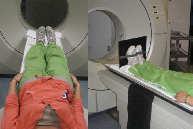
Patient Position
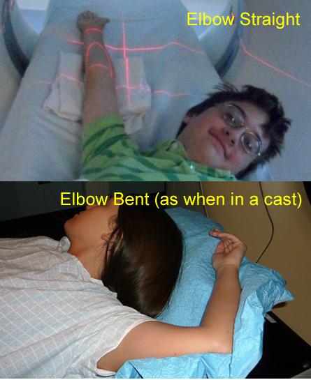
- Supine
- Affected arm raised above head
- Elbow extended (if possible), palm up (if possible)
- Try to position elbow close to the table's center
- Contralateral arm down by the side
Scan Parameters
- SFOV: Small
- kV: 120
- mAs: 150
Reconstruct
- 0.625/0.3 mm Bone
- 2/2 mm Soft Tissue
Coverage
- Humeral shaft to radial shaft (distal to radial tubercle)
DFOV
- Width of anatomy
Straight Elbow: Three reformats
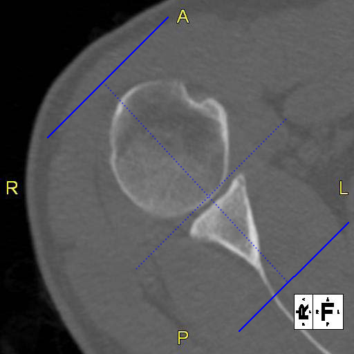
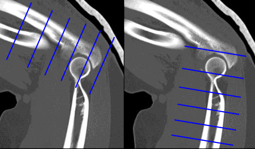
Straight Elbow -- Axial Reformats
- Parallel to line from capitulum to trochlea (distal humerus)
- 0.8/1.5 mm Bone
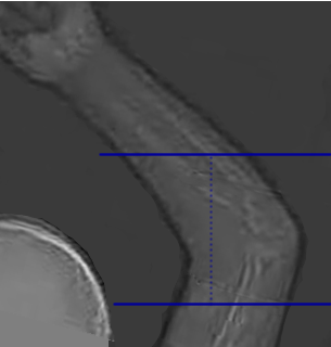
Straight Elbow -- Coronal Reformats
- Prescribe coronal plane off of axial image at level of epicondyles, parallel to inter-epicondylar line
- Orient so humerus is up and forearm is down
- 0.8/1.5 mm Bone
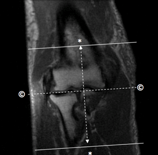
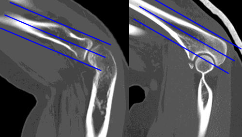
Straight Elbow -- Sagittal Reformats
- Prescribe sagittal plane off of axial image at level of epicondyles, perpendicular to coronal plane
- Orient so humerus is up and forearm is down
- 0.8/1.5 mm Bone
Bent Elbow: Six reformats
Bent Elbow -- Axial Reformats
-
Prescribe axial planes off of a sagittal image;
- Perpendicular to forearm
- Perpendicular to humerus
- 0.8/1.5 mm Bone
Bent Elbow -- Forearm Coronal Reformats
-
Prescribe coronal planes off of sagittal images;
- Perpendicular to radius
- Perpendicular to ulna
- Orient so humerus is up and forearm is down
- 0.8/1.5 mm Bone
Bent Elbow -- Humeral Reformats
-
Prescribe humeral planes off of humeral axial image at level of epicondyles;
- Humeral Coronals: parallel to inter-epicondylar line
- Humeral Sagittals: Perpendicular to inter-epicondylar line
- Orient so humerus is up and forearm is down
- 0.8/1.5 mm Bone
Send Only These...
Straight Elbow
- Scouts
- Source Bone Reconstructions
- Source Soft Tissue Reconstructions
- Axial Reformats
- Coronal Reformats
- Sagittal Reformats
Bent Elbow
- Scouts
- Source Bone Reconstructions
- Source Soft Tissue Reconstructions
- Forearm Axial Reformats
- Humerus Axial Reformats
- Radius Coronal Reformats
- Ulna Coronal Reformats
- Humerus Coronal Reformats
- Humerus Sagittal Reformats
Wrist CT Protocol
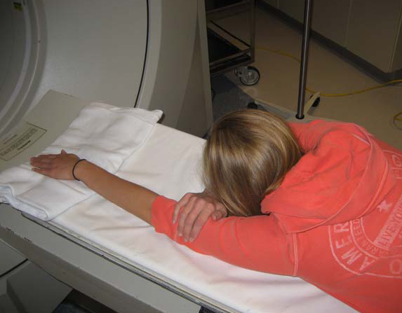
Patient Position
- Prone
- Affected arm over head ("Mighty Mouse" position)
- Arm as straight as possible; palm facing down; wrist centered in gantry
- Contralateral arm by head or at the side
Scan Parameters
- SFOV: Small
- kV: 120
- mAs: 150
Reconstruct
- 0.625/0.3 mm Bone
- 2/2 mm Soft Tissue
Coverage
- Wrist: From the distal radial diaphysis to the third metacarpal base
- Hand: From just proximal to the distal radioulnar joint to include the entire hand
DFOV
- Width of anatomy
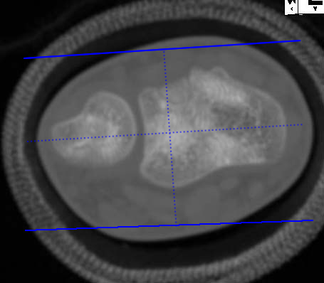
Axial Reformats
- Parallel to distal radius
- 0.8/1.5 mm Bone
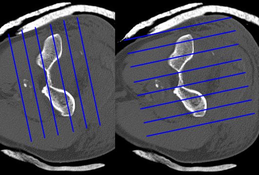
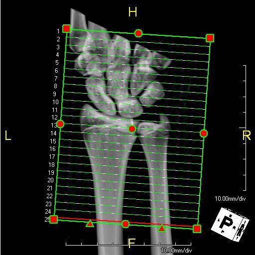
Coronal Reformats
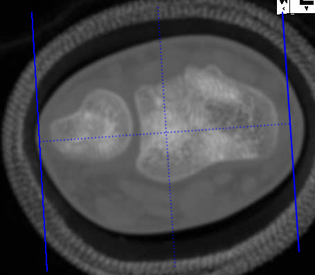
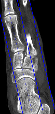
- Prescribe coronal plane off of axial image parallel to line drawn from ulnar styloid to radial styloid.
- Orient so that hand is up and forearm is down
- 0.8/1.5 mm Bone
Sagittal Reformats
- Prescribe sagittal plane off of axial image perpendicular to coronal plane.
- Orient so that hand is up and forearm is down
- 0.8/1.5 mm Bone
Send Only These...
- Scouts
- Source Bone Reconstructions
- Source Soft Tissue Reconstructions
- Axial Reformats
- Coronal Reformats
- Sagittal Reformats
Ankle/Foot CT Protocol
Patient Position
- Supine
- Feet together, centered in scanner; toes pointing straight up; in most cases scan both feet together
- Use foot holder, if available
Scan Parameters
- SFOV: Small
- kV: 120
- mAs: 150
Reconstruct
- 0.625/0.3 mm Bone
- 2/2 mm Soft Tissue
Coverage
- Ankle and Foot: Distal tibial metadiaphysis to include the whole foot
DFOV
- Side of interest only
Ankle/Hindfoot: Three reformats
Ankle/Hindfoot -- Axial Reformats
- Parallel to long axis of calcaneus
- Orient so toes are up and heel is down
- 0.8/1.5 mm Bone
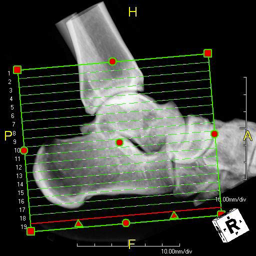
Ankle/Hindfoot -- Coronal Reformats
- Prescribe coronal plane off of axial image at level of distal tib-fib joint, bisecting the tibia and fibula
- Orient so shin is up and foot is down
- 0.8/1.5 mm Bone
Ankle/Hindfoot -- Sagittal Reformats
- Prescribe sagittal plane off of same axial image as coronals, pendicular to coronal plane
- Orient so shin is up and foot is down
- 0.8/1.5 mm Bone
Foot/Forefoot: Three reformats
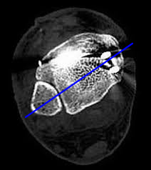
Foot/Forefoot -- Axial Reformats
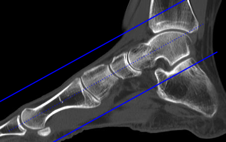
- Prescribe axial plane off of a sagittal image showing most of first MT, parallel to first MT
- Orient so toes are up and heel is down
- 0.8/1.5 mm Bone
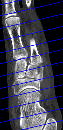
Foot/Forefoot -- Sagittal Reformats
- Prescribe sagittal plane off of a reformatted axial image showing entire first MT, parallel to first MT
- Orient so dorsum of foot is up and plantar surface is down
- 0.8/1.5 mm Bone
Foot/Forefoot -- Coronal Reformats
- Prescribe coronal plane off of the same reformatted axial image as sagittals, perpendicular to sagittals
- Orient so dorsum of foot is up and plantar surface is down
- 0.8/1.5 mm Bone
Send Only These...
Ankle/Hindfoot
- Scouts
- Source Bone Reconstructions
- Source Soft Tissue Reconstructions
- Axial Reformats
- Coronal Reformats
- Sagittal Reformats
Foot/Forefoot
- Scouts
- Source Bone Reconstructions
- Source Soft Tissue Reconstructions
- Axial Reformats
- Coronal Reformats
- Sagittal Reformats